You know the routine with dental X-rays. Bite down on that uncomfortable plastic, hold still, and get a flat picture of your teeth. But what if that simple image is missing the full story? A 3D dental scan creates a complete, high-resolution map of your entire oral structure—teeth, jawbone, sinuses, and airway—in a single 20-second rotation. Unlike flat X-rays, this detailed view shows us how everything is connected. At Primary Integrative Dentistry in Brentwood, Los Angeles, we use this advanced CBCT scanning with AI diagnostics to spot potential issues that standard imaging simply can’t see.
Make an appointment
What is a CBCT 3D Dental Scan?
A CBCT dental scan (cone beam computed tomography) is an advanced imaging technology that uses a cone-shaped X-ray beam to capture hundreds of images as it rotates around your head. Primary Integrative Dentistry uses CBCT scanning to produce a comprehensive 3D reconstruction of teeth, bone, nerves, soft tissue, and airway passages in a single scan lasting approximately 20 seconds.
A computer processes these images into a three-dimensional model that your clinician can rotate, slice, and zoom into from any angle. This means Dr. Tzur Gabi can examine the precise thickness of your jawbone before placing an implant, trace the path of a nerve before a surgical procedure, or measure the diameter of your airway to screen for sleep-disordered breathing.
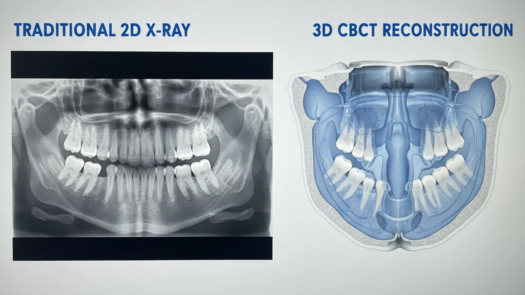
The technology differs fundamentally from the flat, overlapping images produced by traditional dental X-rays. Where a standard X-ray compresses three-dimensional structures into a single plane, CBCT preserves spatial relationships and depth. The result is diagnostic precision that changes treatment planning from educated guesswork into data-driven decision-making.
How Does the Technology Work?
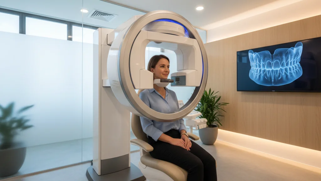
If you’ve ever had a traditional dental X-ray, you know the routine: biting down on an uncomfortable plastic film while a technician takes multiple shots. A CBCT scan is a much simpler and more comfortable process. You’ll stand or sit still for just a few seconds—typically less than 20 for a full scan—while a machine arm makes a single, quiet rotation around your head. The entire scan is completely painless. During that one pass, the cone-shaped X-ray beam captures hundreds of individual 2D images from every possible angle, creating a complete dataset without you having to move or readjust. It’s a bit like a high-speed photoshoot for your jaw.
The real magic happens in the software. A powerful computer processes all those flat pictures, stitching them together to create an incredibly detailed, three-dimensional digital model of your anatomy. This isn’t just a picture of your teeth; it’s a complete map showing your bones, soft tissues, nerve pathways, and even your airway in their exact spatial relationship to one another. This level of detail is what allows us to see the full picture of your oral health. At Primary Integrative Dentistry, we use this advanced 3-D scanning technology to move beyond just treating symptoms, identifying the true root cause of issues and ensuring our treatment plans are precise and effective.
3D Dental Scans vs. X-Rays: What’s the Difference?
Traditional dental X-rays have served dentistry well for over a century, but they have inherent limitations. A standard periapical or panoramic X-ray produces a flat, two-dimensional image where structures overlap and important details can hide behind other anatomy. A 3D dental scan eliminates these blind spots.
Here is how the two technologies compare:
| Feature | Traditional 2D X-Ray | 3D CBCT Scan |
|---|---|---|
| Image type | Flat, single plane | Full 3D volume |
| Scan time | 1-5 seconds per image | 15-20 seconds total |
| Anatomy captured | Teeth and surrounding bone | Teeth, jaw, sinuses, airway, nerves, TMJ |
| Overlapping structures | Common, can hide pathology | Eliminated through 3D slicing |
| Radiation dose | Very low per image | Low; comparable to a set of full-mouth X-rays |
| Measurement accuracy | Limited, prone to distortion | Sub-millimeter precision |
| Airway visualization | Not possible | Full volumetric airway mapping |
The practical impact is significant. Research published in the Journal of Endodontics found that CBCT imaging detected up to 34% more periapical lesions (infections at tooth roots) than conventional radiography. For patients, that translates to catching problems earlier and avoiding more invasive treatments down the line.
At Primary Integrative Dentistry, the CBCT scan is just the starting point. Every scan is also analyzed by Overjet, an FDA-cleared AI diagnostic platform that highlights areas of concern with color-coded overlays, quantifies bone levels with sub-millimeter precision, and flags early-stage decay that even experienced clinicians might miss on a first pass.
Make an appointment
When Are Traditional X-Rays a Better Fit?
While 3D scanning provides incredible diagnostic detail, traditional 2D X-rays still play a role in modern dentistry. For a routine check-up where the goal is simply to spot surface-level cavities, a standard bitewing X-ray is often the most efficient tool. It’s fast, uses a very low radiation dose, and is perfectly suited for that one specific job. But its usefulness ends where complexity begins. When we need to investigate the root cause of pain, precisely plan for a dental implant, or evaluate the health of your jawbone, a flat image simply doesn’t give us the whole story. Relying on it for complex diagnostics is like trying to navigate a city with a map that’s missing half its streets.
Common Types of 2D X-Rays
You’ve probably had a few different types of 2D X-rays during past dental appointments. The most common are bitewings, which give us a clear view of the crowns of your back teeth to find decay hiding between them. Periapical X-rays focus on a single tooth, showing everything from the crown to the tip of the root, helping diagnose issues in the surrounding bone. Then there’s the panoramic X-ray, which creates one wide, flat image of your entire mouth, jaws, and sinuses. While helpful for a general overview, each of these flattens your anatomy into a single plane. This compression layers structures on top of each other, potentially hiding tiny fractures or infections—which is exactly why our practice relies on 3-D scanning to get the complete picture.
What a 3D Dental Scan Reveals About Your Teeth
A 3D dental scan provides a level of detail about tooth structure that two-dimensional imaging simply cannot match. Primary Integrative Dentistry uses CBCT technology to evaluate teeth from every possible angle, revealing conditions that would otherwise require multiple appointments and imaging sessions to diagnose.
Spotting Hidden Cracks and Decay
Hairline cracks in teeth are notoriously difficult to detect on standard X-rays. CBCT scanning reveals vertical root fractures, craze lines, and internal resorption that can cause persistent pain without an obvious visible cause. Identifying these fractures early prevents unnecessary root canal treatments on teeth that actually need extraction or a different intervention.
Mapping Complex Root Canals
Teeth often have more root canals than textbooks suggest. Maxillary molars, for example, can have four or five canals instead of the expected three. A 3D scan maps every canal before treatment begins, reducing the risk of missed canals that lead to persistent infection and retreatment.
Detecting Early Infection and Bone Loss
Periapical abscesses (infections at the tip of a tooth root) appear on CBCT scans before they become visible on traditional X-rays. The 3D view also reveals the exact size, shape, and location of the infection, helping Dr. Gabi determine whether the tooth can be saved through regenerative endodontics and other root canal alternatives or whether extraction is the better path forward.
Pinpointing Impacted Teeth
For wisdom teeth or other impacted teeth, CBCT scanning shows the precise relationship between the impacted tooth and surrounding structures, including the inferior alveolar nerve, adjacent tooth roots, and the maxillary sinus. This information is critical for planning safe surgical extractions.
Creating Better-Fitting Appliances
Whether it’s a night guard, a retainer, or a surgical guide for a dental implant, a perfect fit is non-negotiable for both comfort and effectiveness. A 3D dental scan creates a precise digital model of your teeth and jaw, providing sub-millimeter data that a physical impression simply can’t capture. This level of detail allows for the creation of custom appliances that fit perfectly from day one. It transforms the process from educated guesswork into data-driven design, ensuring your appliance is not only effective but also comfortable enough for consistent use.
Beyond Teeth: What a Scan Shows About Your Jaw
Jawbone health determines the foundation of your entire oral system. A CBCT scan provides Primary Integrative Dentistry with precise measurements of bone density, height, width, and quality that guide treatment decisions for implants, prosthetics, and structural rehabilitation.
Planning for a Perfect Dental Implant
Dental implant success depends on placing the implant in bone that is dense enough and thick enough to support it. A 3D dental scan measures bone dimensions to the fraction of a millimeter, allowing Dr. Tzur Gabi, a board-certified prosthodontist, to select the correct implant size, angle, and position before surgery begins. This precision is particularly valuable for complex cases like zirconia implants or full-arch reconstructions.
Getting a Clearer View of Your TMJ
The temporomandibular joint (TMJ) is a complex structure that traditional X-rays capture poorly. CBCT scanning reveals the shape and position of the condyle, disc space, and articular eminence in three dimensions. For patients experiencing jaw pain, clicking, or limited opening, this imaging provides the diagnostic clarity needed to distinguish between muscular TMJ disorders and structural joint pathology.
Do You Need a Bone Graft?
Patients who have experienced bone loss from periodontal disease, tooth extraction, or trauma need accurate measurements before bone grafting procedures. The 3D scan shows exactly how much bone has been lost and where grafting material needs to be placed, improving surgical outcomes and reducing recovery time.
Screening for Cysts and Abnormalities
Cysts, tumors, and other jaw pathologies are visible on CBCT scans at much earlier stages than on traditional imaging. The three-dimensional view reveals the extent of the lesion and its relationship to surrounding structures, providing critical information for treatment planning.
Planning for Reconstructive Surgery
Successful oral surgery isn’t just about what happens in the chair; it’s about the meticulous planning that happens long before. When you’re rebuilding a smile with a dental implant or full-arch reconstruction, a 3D scan provides the essential blueprint. It offers precise measurements of your jawbone’s density and dimensions, allowing your dentist to digitally map the entire surgery for perfect placement. If the scan reveals bone loss from past extractions or disease, it shows exactly where a graft is needed to create a stable foundation. This data-driven approach ensures every step is engineered for a successful, long-lasting outcome.
The scan also acts as a critical safety check. Cysts, tumors, and other abnormalities are visible on a CBCT scan at much earlier stages than on traditional imaging, revealing their exact size and location relative to nerves and sinuses. This comprehensive view is vital for creating a safe surgical plan that avoids surprises and protects surrounding healthy structures. It’s a key part of our integrated approach to all dental services, ensuring your procedure is both effective and safe.
Can a Dental Scan Help with Sleep Apnea?
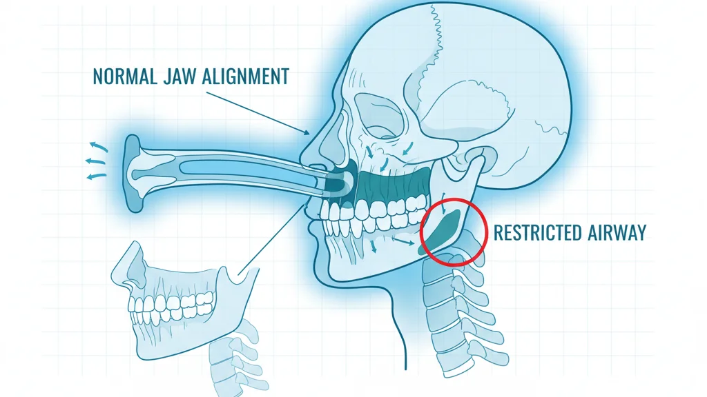
One of the most clinically significant capabilities of a 3D dental scan is volumetric airway analysis. Primary Integrative Dentistry uses CBCT imaging to measure the size, shape, and narrowest points of your airway, providing objective data that connects your jaw structure to your breathing and sleep quality.
A CBCT scan captures the full airway from the nasal passages through the pharynx, measuring the cross-sectional area at every level. For patients with obstructive sleep apnea or sleep-disordered breathing, this data reveals exactly where and why the airway narrows during relaxation.
Analyzing Your Airway for Blockages
- Minimum cross-sectional area of the airway, measured in square millimeters. An airway below 110mm2 is considered at elevated risk for obstruction during sleep.
- Airway volume from the hard palate to the base of the epiglottis, providing an overall assessment of breathing capacity.
- Location of maximum constriction, which determines whether the obstruction is at the level of the soft palate, tongue base, or lateral pharyngeal walls.
- Jaw position and retrognathia, where a recessed lower jaw pushes the tongue backward and compresses the airway.
- Nasal passage patency, identifying deviated septum, turbinate hypertrophy, or sinus blockages that contribute to mouth breathing.
This airway data is essential for CPAP refugees, patients who have been diagnosed with sleep apnea but cannot tolerate a CPAP machine. By identifying the anatomical cause of obstruction, Primary Integrative Dentistry can determine whether an oral appliance, myofunctional therapy, or a referral for surgical intervention is the most appropriate treatment path.
Is a 3D Dental Scan Right for You?
Not every dental visit requires CBCT imaging, but the technology is essential for accurate diagnosis and treatment planning in several common clinical scenarios. Primary Integrative Dentistry recommends 3D dental scans for patients in the following situations:
- Dental implant candidates. Precise bone measurements are required before any implant placement.
- Complex root canal cases. Multi-rooted teeth, retreatments, or suspected vertical root fractures benefit from 3D visualization.
- Impacted wisdom teeth. Mapping the relationship between impacted teeth and the inferior alveolar nerve prevents nerve injury during extraction.
- TMJ and jaw pain. Structural assessment of the temporomandibular joint requires three-dimensional imaging.
- Sleep apnea screening. Volumetric airway analysis identifies anatomical risk factors for obstructive sleep apnea.
- Orthodontic evaluation. Treatment planning for aligners, such as Invisalign or NiTime, benefits from understanding root positions and bone boundaries.
- Unexplained facial pain. CBCT can identify sinus disease, bone pathology, or infections not visible on standard X-rays.
- Sinus evaluation. Dental procedures near the maxillary sinus, such as upper implants, require knowing sinus floor height and membrane thickness.
- New patient comprehensive exams. At Primary Integrative Dentistry, CBCT scanning is part of every new patient visit because it establishes a complete baseline of your oral, structural, and airway health.
The free comprehensive exam at Primary Integrative Dentistry includes a full 360-degree scan, CBCT imaging, digital X-rays, AI-powered analysis, and screening for 99+ health concerns.
Your 3D Dental Scan: What to Expect
The CBCT scanning process at Primary Integrative Dentistry is fast, painless, and non-invasive. Here is what happens during your appointment:
- Preparation. You will remove any jewelry, glasses, or metal accessories from your head and neck area. No special preparation or fasting is required.
- Positioning. You stand or sit in the scanner while a technician adjusts the machine to align with your anatomy. A chin rest and head stabilizer keep you comfortable and still during the scan.
- The scan. The CBCT arm rotates around your head in a single pass lasting approximately 15 to 20 seconds. You will hear a quiet mechanical sound but feel nothing. The radiation dose is low, comparable to a standard set of full-mouth dental X-rays and significantly less than a medical CT scan.
- AI analysis. Your scan data is processed by Overjet AI technology, which automatically analyzes your radiographs for decay, bone loss, periodontal disease, and calculus. This AI analysis provides Dr. Gabi with a second set of eyes that never fatigues and has been trained on millions of dental images.
- Review with your clinician. Dr. Tzur Gabi reviews the 3D images with you on screen, rotating and slicing through the scan to show you exactly what is happening in your teeth, jaw, sinuses, and airway. You see what your clinician sees, in real time.
The entire imaging process takes less than five minutes from start to finish. Results are available immediately because the processing happens on-site.
Make an appointment
The Scan Procedure: Step-by-Step
The idea of a “3D scan” might sound complex, but the actual process is surprisingly simple and quick. The entire experience is designed to be comfortable and efficient, getting you from the chair to a complete picture of your oral health in just a few minutes. There are no tight spaces or loud noises—just a straightforward process that provides an incredible amount of diagnostic information.
Preparing for Your Scan
Before the scan begins, we’ll ask you to remove any metal objects from your head and neck area, like jewelry, glasses, or removable dental appliances. These items can interfere with the X-ray beam and affect the clarity of the final image. The good news is that no other special preparation is needed. You don’t have to fast or change your routine; you can come straight from work or home and be ready to go.
During the Scan
You’ll either stand or sit in the open-air scanner, and a team member will gently position your head using a comfortable chin rest and stabilizers. This simply ensures you stay still, which is key to getting a crisp, clear image. Once you’re ready, the scanner’s arm will make a single, smooth rotation around your head that takes about 15 to 20 seconds. You’ll hear a quiet mechanical whirring, but you won’t feel a thing. The radiation exposure is minimal—comparable to a standard set of full-mouth dental X-rays and far less than a medical CT scan.
Who Interprets Your Scan Results?
Your scan isn’t sent off to a lab for a stranger to read. At Primary Integrative Dentistry, your results are interpreted with you, not just for you. Dr. Tzur Gabi will sit down with you and pull up the 3D model on a large screen right in the treatment room. Together, you’ll explore the intricate details of your anatomy as he rotates and slices through the images, pointing out exactly what he sees in your teeth, jaw, sinuses, and airway. This collaborative review is a core part of our wholistic approach, turning a diagnostic tool into an educational experience that empowers you to understand the root causes of your health concerns.
Are 3D Dental Scans Safe?
CBCT scanning uses ionizing radiation, but the dose is carefully controlled and significantly lower than medical CT imaging. A typical dental CBCT scan delivers between 20 and 200 microsieverts, depending on the field of view. For context, the average annual background radiation exposure from natural sources is approximately 3,000 microsieverts.
The American Dental Association and the American Academy of Oral and Maxillofacial Radiology both endorse CBCT imaging when the clinical benefit justifies the minimal radiation exposure. At Primary Integrative Dentistry, Dr. Gabi follows the ALARA principle (As Low As Reasonably Achievable), recommending CBCT only when the diagnostic information it provides cannot be obtained through lower-dose imaging.
For pregnant patients, CBCT scanning is generally deferred unless there is an emergency clinical need, consistent with standard radiation safety protocols across all medical imaging.
Understanding Radiation Exposure
It’s completely normal to have questions about radiation, so it’s helpful to put the dose from a dental CBCT scan into context. The exposure is extremely low and used very intentionally. A single scan delivers a radiation dose between 20 and 200 microsieverts, while the average person is exposed to about 3,000 microsieverts every year from natural background sources. This means a focused dental scan is a small fraction of the exposure you get from simply living on Earth for a year. It’s also significantly less radiation than a conventional medical CT scan, making it a safer, more targeted option for detailed dental and facial imaging.
Special Considerations for Children
For our younger patients, we take an even more cautious approach. Because children’s developing tissues are more sensitive, we only recommend a 3D scan when it is absolutely necessary for an accurate diagnosis or safe treatment plan—for instance, with complex orthodontic cases or impacted teeth. We adhere strictly to safety protocols, using the smallest possible field of view and the lowest effective exposure settings to capture only the required information. This careful approach ensures we gain the diagnostic clarity needed for the best possible care while always prioritizing your child’s long-term health and well-being.
Weighing the Benefits Against the Risks
At Primary Integrative Dentistry, every decision is guided by a patient-first philosophy. We follow a principle known as ALARA, which stands for “As Low As Reasonably Achievable.” This means Dr. Gabi will only recommend a 3D scan when the detailed diagnostic information it provides is essential for your care and cannot be obtained with lower-dose imaging. The American Dental Association supports this approach, endorsing CBCT when the clinical benefits justify the minimal exposure. This tool allows us to diagnose issues with incredible precision, which ultimately leads to more effective, less invasive treatments—a core tenet of our wholistic approach to dentistry.
How AI Gives You a Smarter Diagnosis
Primary Integrative Dentistry combines CBCT imaging with AI-powered diagnostics through Overjet, an FDA-cleared platform that adds a layer of machine intelligence to every scan. This combination represents one of the most advanced diagnostic workflows available in dentistry today.
Overjet analyzes your dental radiographs in real time and performs several critical functions:
- Decay detection and outlining. The AI identifies and outlines areas of tooth decay with 93% accuracy, catching early-stage cavities that might not be visible to the naked eye.
- Bone level quantification. Instead of subjective visual assessment, Overjet provides precise millimeter measurements of bone levels around each tooth, essential for tracking periodontal disease progression.
- Periodontal disease identification. Pattern recognition trained on millions of images flags gum disease indicators before they progress to tooth loss.
- Calculus detection. Tartar deposits that need professional removal are automatically highlighted.
When combined with the three-dimensional anatomical data from CBCT, the result is a comprehensive diagnostic picture that connects your tooth-level findings to your jawbone health, sinus condition, and airway function. This is the foundation of the Primary iD health scoring system, which tracks your wellness across five dimensions: Oral Health, Sleep and Lifestyle, Nutrition, Genetics and Microbiome, and Airway Health.
Frequently Asked Questions About 3D Dental Scans
How much does a 3D dental scan cost? The cost of a CBCT scan varies depending on the area being imaged and your insurance coverage. Many dental insurance plans cover CBCT imaging when medically necessary. At Primary Integrative Dentistry, the free new patient comprehensive exam includes CBCT imaging, digital X-rays, a 360-degree scan, and AI-powered analysis.
How long does a CBCT scan take? The scan itself takes 15 to 20 seconds. The entire imaging appointment, including positioning and review, takes less than five minutes.
Is a 3D dental scan painful? No. CBCT scanning is completely painless and non-invasive. You simply stand still while the scanner rotates around your head.
How often should I get a 3D dental scan? CBCT imaging is prescribed based on clinical need, not on a fixed schedule. Your dentist will recommend a scan when the diagnostic benefit justifies it, such as before implant placement, to evaluate unexplained symptoms, or to establish a baseline at your first visit.
Can a CBCT scan detect cancer? CBCT scanning can reveal abnormal growths, cysts, and tumors in the jaw and surrounding structures. While CBCT is not a cancer screening tool, incidental findings of suspicious lesions do occur and are referred for appropriate follow-up.
What is the difference between a CBCT scan and a medical CT scan? CBCT uses a cone-shaped beam and a single rotation, producing lower radiation doses and focusing specifically on the head and neck region. Medical CT scanners use a fan-shaped beam with multiple rotations and are designed for whole-body imaging at higher radiation doses.
Does insurance cover 3D dental scans? Many dental insurance plans cover CBCT imaging when it is medically necessary for diagnosis or treatment planning. Primary Integrative Dentistry accepts most insurance plans and can verify your coverage before your appointment.
Can children get CBCT scans? Yes, when clinically indicated. CBCT is used in pediatric dentistry for evaluating impacted teeth, airway assessment, and orthodontic planning. The radiation dose is adjusted for smaller patients.
How Much Does a 3D Dental Scan Cost?
When considering a 3D dental scan, one of the first questions is usually about the price. Generally, you can expect a CBCT scan to cost between $350 and $600. This investment provides an unparalleled level of diagnostic detail that can help you avoid more complex and costly treatments in the future by catching issues early. At Primary Integrative Dentistry, we believe this foundational diagnostic step is so critical to understanding your overall health that we include a 3D CBCT scan as part of our comprehensive new patient exam, which is offered at no cost. This allows us to establish a complete and accurate baseline of your oral and airway health from day one, without cost being a barrier.
Factors That Influence the Price
Several factors can influence the out-of-pocket cost of a CBCT scan if it’s not part of an initial exam. The clinic’s geographic location plays a role, as costs can vary between major metropolitan areas and smaller towns. The specific reason for the scan also matters; for example, imaging for a single dental implant may differ in scope from a scan needed for a full-mouth reconstruction or complex oral surgery. Finally, the type of insurance coverage you have will significantly impact your final cost, as different plans offer varying levels of reimbursement for advanced diagnostic imaging.
Will Insurance Cover a 3D Scan?
The good news is that many dental insurance plans do cover CBCT scans, especially when they are deemed “medically necessary.” This typically includes situations where the scan is essential for accurate diagnosis or safe treatment planning, such as mapping nerve pathways before an extraction, measuring bone density for implants, or evaluating a complex infection. It’s not usually covered for purely elective or cosmetic reasons. Our team at Primary Integrative Dentistry is experienced in working with insurance providers and can help you understand your benefits and submit the necessary documentation to maximize your coverage for any recommended services.
Key Takeaways
- See the complete picture of your oral health: A 3D CBCT scan provides a detailed map of your teeth, jaw, sinuses, and airway, revealing hidden issues like hairline cracks or early infections that traditional flat X-rays can’t show.
- Connect your oral health to your overall wellness: This advanced imaging provides crucial data for planning dental implants, assessing your jaw joint, and even analyzing your airway to screen for potential sleep-disordered breathing.
- Make informed decisions with a smarter diagnosis: The scan itself is fast and comfortable, using a low radiation dose to produce immediate results that, when combined with AI analysis, give you and your clinician a precise, data-driven foundation for your treatment plan.